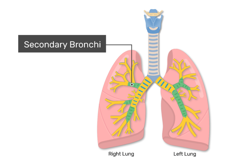
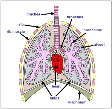
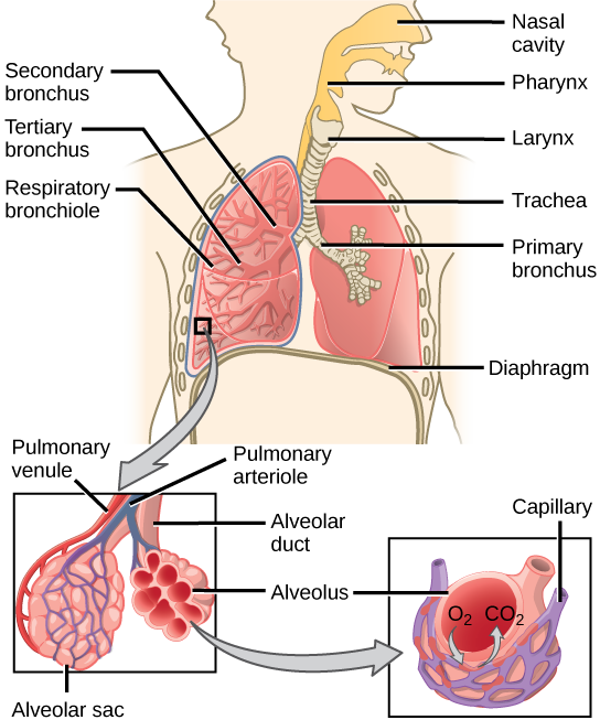
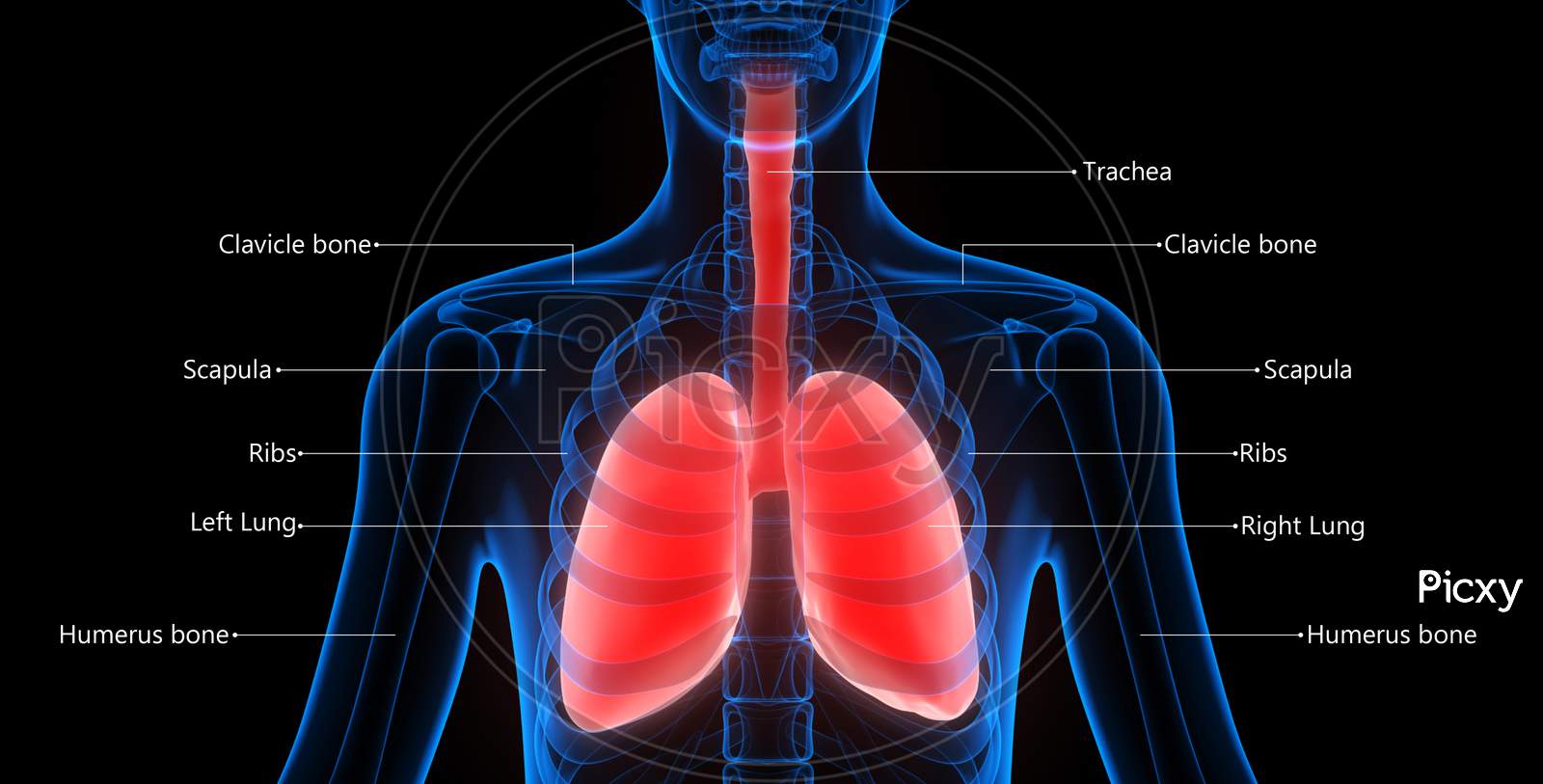
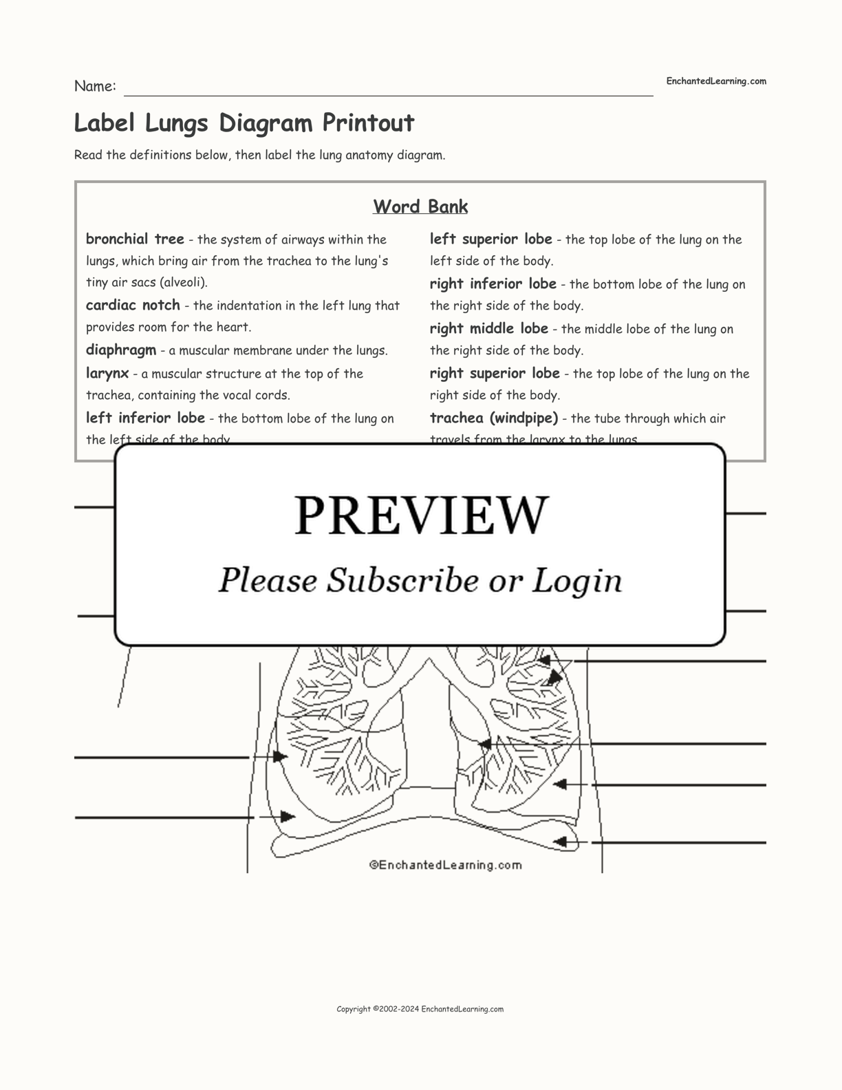
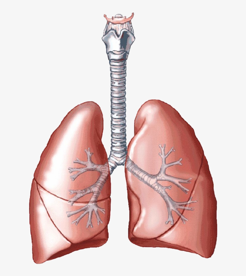
45 diagram of the lungs with labels
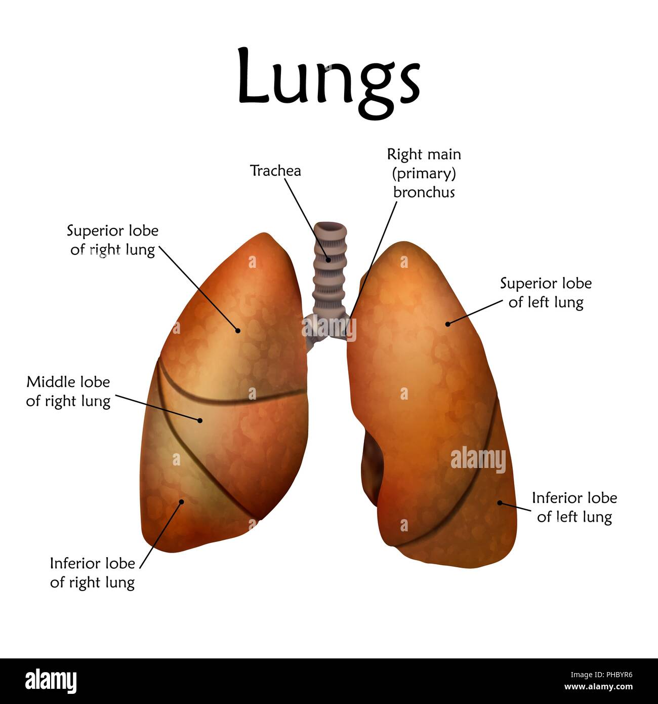
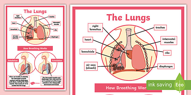
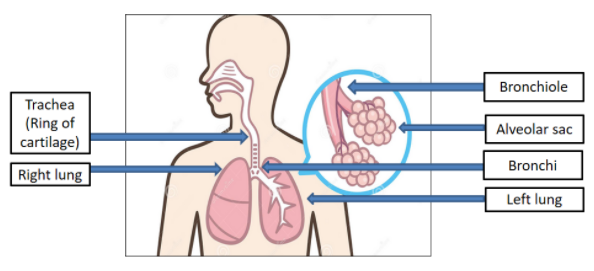
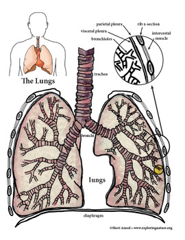
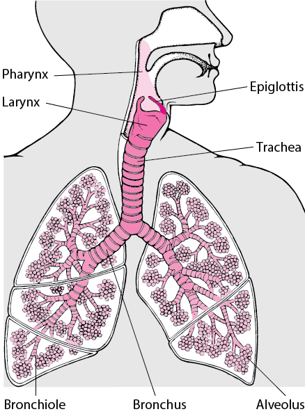
Labeled diagram of the lungs/respiratory system. - SERC Jan 10, 2014 · Labeled diagram of the lungs/respiratory system. Image 37789 is a 1125 by 1408 pixel PNG. Uploaded: Jan10 14. Last Modified: 2014-01-10 12:15:34. Permanent URL: . The file is referred to in 1 page. Airborne Microbes. Lung Diagram Labeled | EdrawMax Template Mar 02, 2022 · Labeled The following is the elaborated diagram of human lungs. In the following lung labeled diagram, we have shown Thyroid cartilage, Cricoid Cartilage, Tracheal Cartilage, Apex, Left Upper Lobe, Hilum, Left Bronchus, Oblique Fissure, Bronchioles, Left Lower lobe, Base of lung, cardiac notch, right lower lobe, oblique fissure, right middle lobe, horizontal fissure, right bronchus, right upper lobe, and trachea.
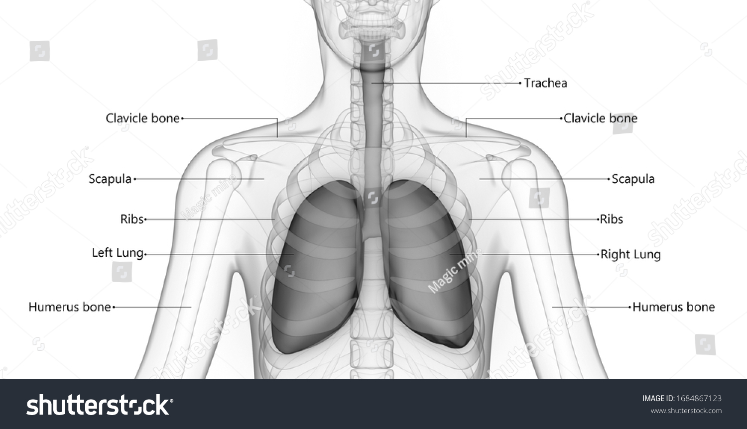
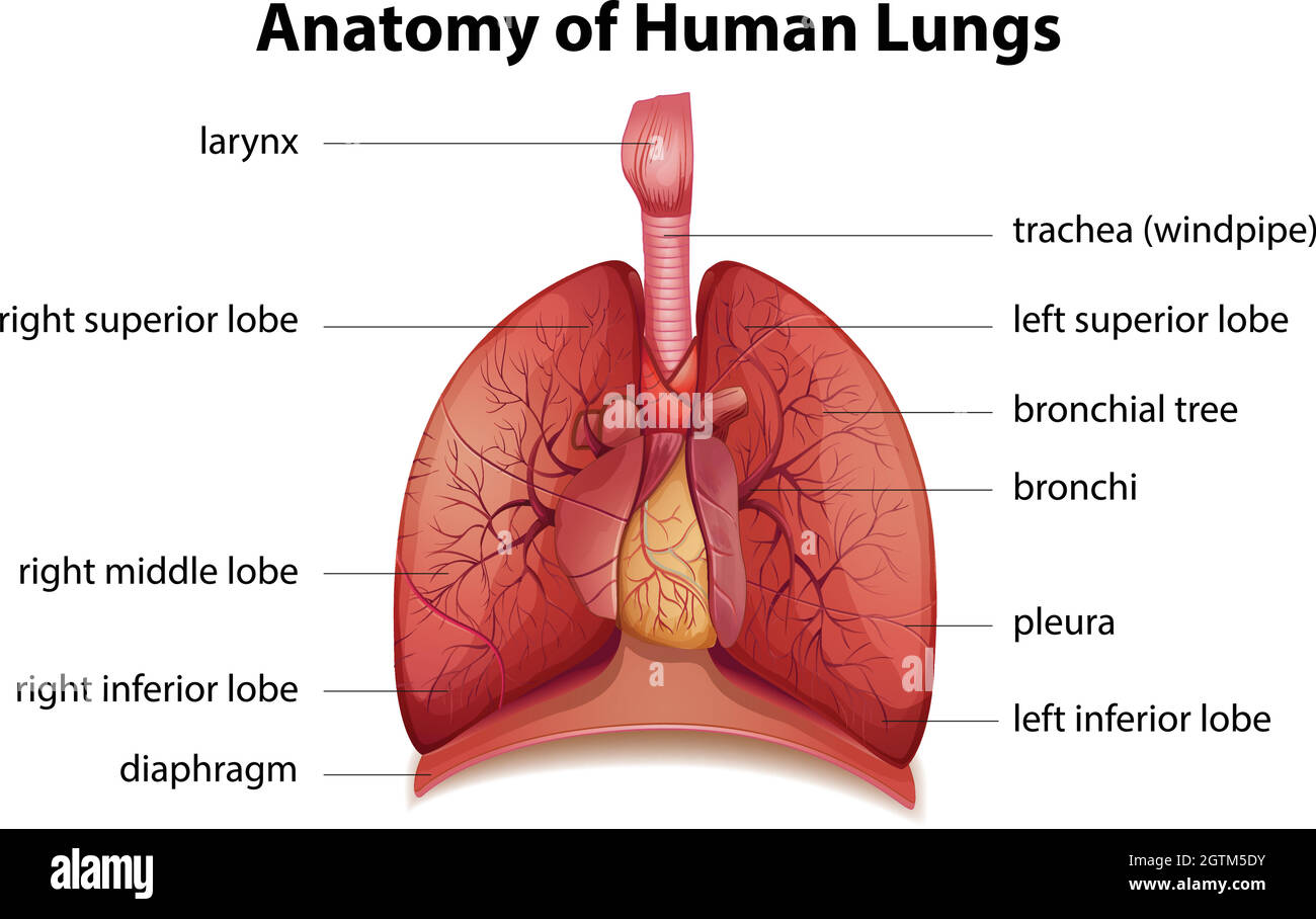
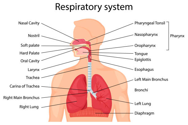
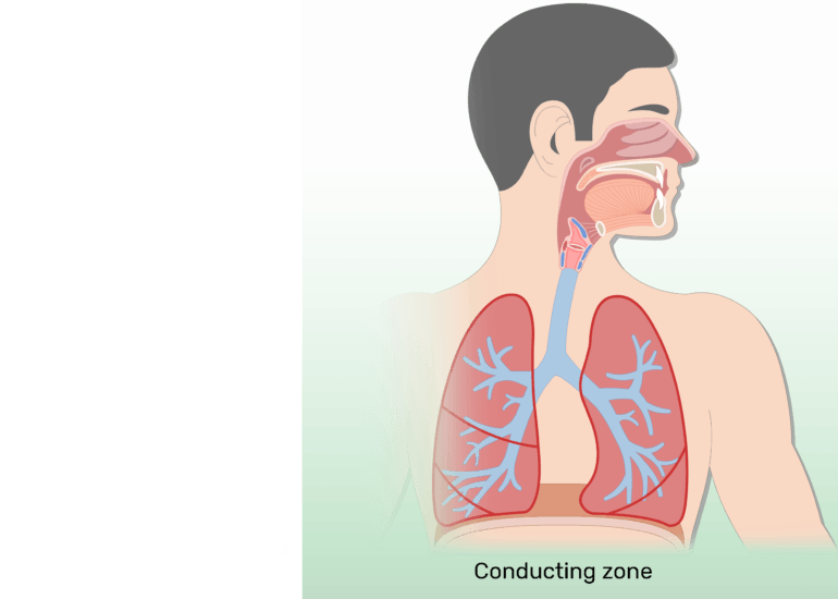
Lungs Diagram - Human Lungs Anatomy - BYJUS Lungs Diagram in Human Body. Humans have a right and a left lung positioned in the chest cavity. Jointly, the lungs inhabit most of the intrathoracic space. Lungs are responsible for adding oxygen and removing carbon dioxide from the blood, thus serving as a gas-exchanging structure for respiration. Each of the lungs in humans is encased in pleura – a thin membranous sac, and each is linked with the trachea by its main bronchus, with that of the heart by pulmonary arteries.
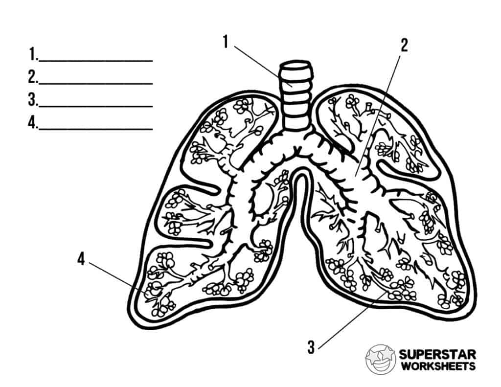
Diagram of the lungs with labels
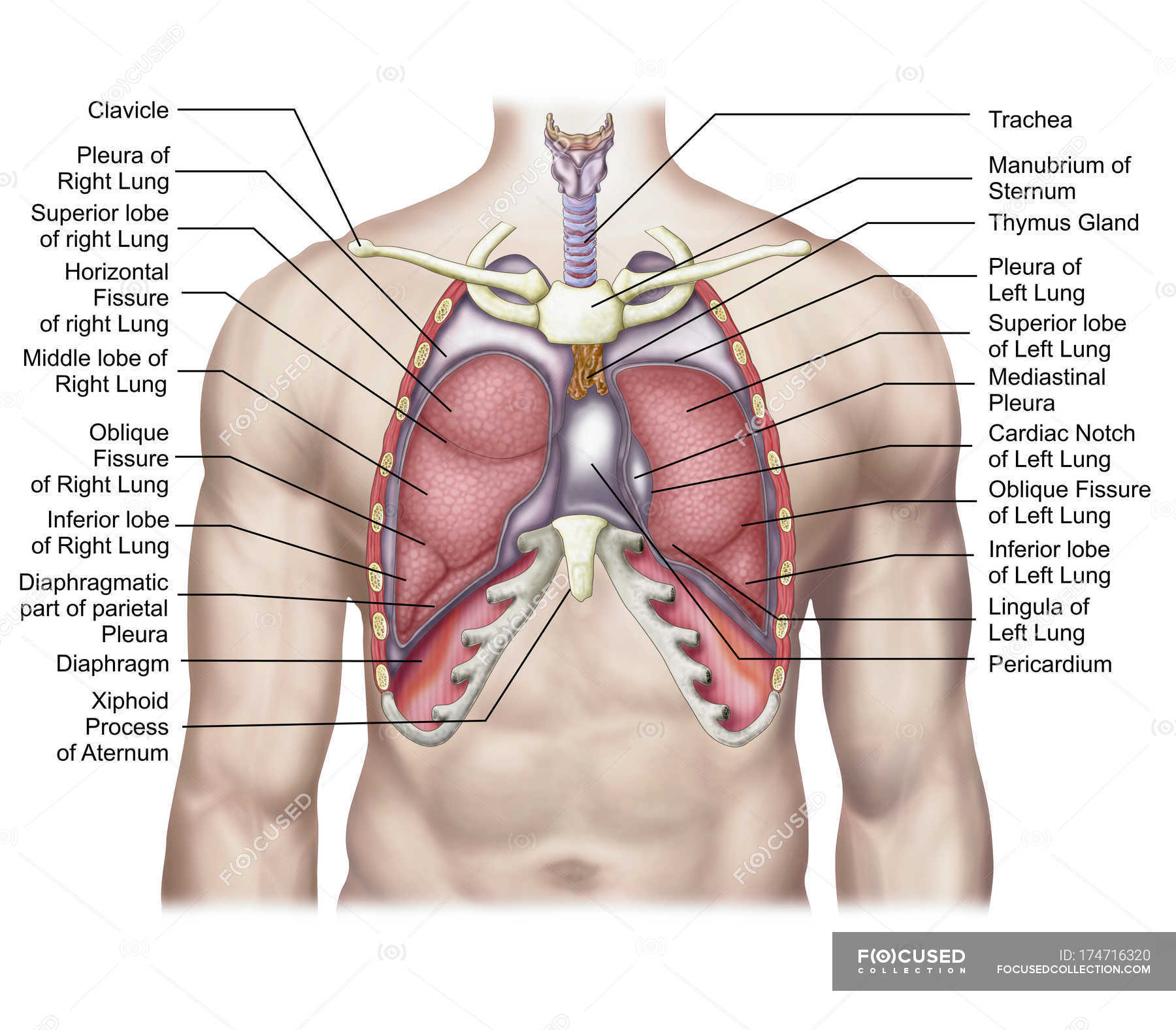
Lungs Diagram - Structure, Working, Importance and Facts Structure and Simple Lungs Diagram. One on each side of the chest, a human body has two lungs. Between the lungs is where the heart is located. The lobes of the right lung are three rounded sections. The left lung is divided into two lobes. The diaphragm is a strong muscle sheet that rests at the base of each lung. Labeled Diagram of the Human Lungs - Bodytomy Given below is a labeled diagram of the human lungs followed by a brief account of the different parts of the lungs and their functions. Each lung is enclosed inside a sac called pleura, which is a double-membrane structure formed by a smooth membrane called serous membrane. Lung Anatomy, Function, and Diagrams - Healthline Dec 14, 2018 · The lungs begin at the bottom of your trachea (windpipe). The trachea is a tube that carries the air in and out of your lungs. Each lung has a tube called a bronchus that connects to the trachea ...
Diagram of the lungs with labels. Lung Anatomy, Function, and Diagrams - Healthline Dec 14, 2018 · The lungs begin at the bottom of your trachea (windpipe). The trachea is a tube that carries the air in and out of your lungs. Each lung has a tube called a bronchus that connects to the trachea ... Labeled Diagram of the Human Lungs - Bodytomy Given below is a labeled diagram of the human lungs followed by a brief account of the different parts of the lungs and their functions. Each lung is enclosed inside a sac called pleura, which is a double-membrane structure formed by a smooth membrane called serous membrane. Lungs Diagram - Structure, Working, Importance and Facts Structure and Simple Lungs Diagram. One on each side of the chest, a human body has two lungs. Between the lungs is where the heart is located. The lobes of the right lung are three rounded sections. The left lung is divided into two lobes. The diaphragm is a strong muscle sheet that rests at the base of each lung.










Post a Comment for "45 diagram of the lungs with labels"