39 fluorescent labels and light microscopy
Fluorescence Microscopy vs. Light Microscopy - News-Medical.net This means that fluorescent microscopy uses reflected rather than transmitted light. For example, a commonly used label is green fluorescent protein (GFP), which is excited with blue light and... Researchers demonstrate label-free super-resolution microscopy - Phys.org A newly developed sub-diffraction-limit microscopy approach doesn't require fluorescent labels. The video shows the process of the data evaluation algorithm, retrieving the positions and sizes of...
Immunolabeling for Correlative Light and Electron Microscopy on ... An ideal label for light and electron microscopy would contain both fluorescent dye and a colloidal nanoparticle on a single antibody molecule. Secondary and tertiary label, the colloidal nanoparticles are placed on the primary antibody while the fluorescent dye conjugated to secondary ( D) or tertiary ( E) antibody.

Fluorescent labels and light microscopy
Fluorescent labeling of abundant reactive entities (FLARE) for cleared ... Fluorescence microscopy is a technique that is commonly used in the biomedical sciences. It offers the powerful ability to visualize structures or molecules in three dimensions within biological... Fluorescence Imaging - Teledyne Photometrics Fluorescent molecules (known as fluorophores) are used to label samples, and fluorophores are available that emit light in virtually any color. In a fluorescent microscope, a sample is labeled with a fluorophore, and then a bright light ( excitation light) is used to illuminate the sample, which gives off fluorescence ( emission light ). Dots, Probes and Proteins: Fluorescent Labels for Microscopy and Imaging There are currently 20 AlexaFluor dyes which span the excitation spectrum from 346 nm to 784 nm and are available as labelling kits, or conjugated to primary or secondary antibodies. Thermo Scientific produce their own probes called ' DyLight '. The ten probes which are currently available cover a spectrum from 350 nm to 800 nm.
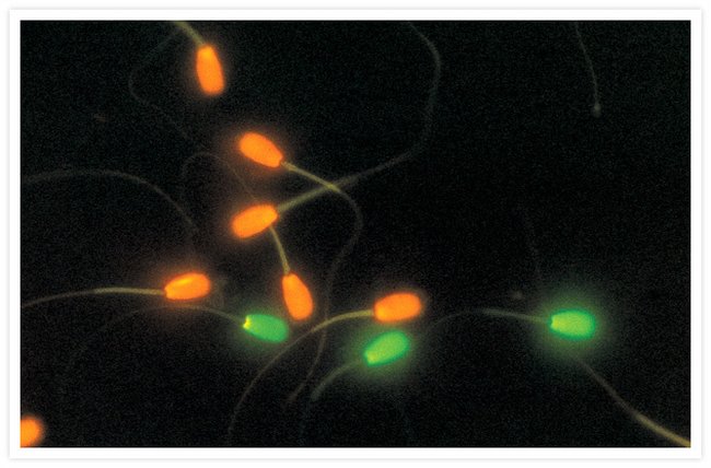
Fluorescent labels and light microscopy. Optical Microscopy Application: Fluorescence | Edmund Optics Optical Microscopy Application: Fluorescence. Fluorescence microscopy is an optical microscopy technique that utilizes fluorescence, which is induced using fluorophores, as opposed to absorption, scatter, or reflection. A fluorophore is a type of fluorescent dye used to mark proteins, tissues, and cells with a fluorescent label for examination ... Light Sheet Fluorescence Microscopy - an overview - ScienceDirect Applications of single-molecule fluorescence microscopy. (A) The photophysical properties of a fluorophore contain information about its position and its state. This allows, for example, tracking molecules, observing conformational and constitutional changes, or following chemical reactions. (B) Examples for applications in biology and chemistry. Fluorescence Microscopy - an overview | ScienceDirect Topics Fluorescence microscopy is a technique whereby fluorescent substances are examined in a microscope. It has a number of advantages over other forms of microscopy, offering high sensitivity and specificity. In fluorescence microscopy, the specimen is illuminated (excited) with light of a relatively short wavelength, usually blue or ultraviolet (UV). Fluorescent tag - Wikipedia S. cerevisiae septins revealed with fluorescent microscopy utilizing fluorescent labeling In molecular biology and biotechnology, a fluorescent tag, also known as a fluorescent label or fluorescent probe, is a molecule that is attached chemically to aid in the detection of a biomolecule such as a protein, antibody, or amino acid.
Fluorescent Labeling - What You Should Know - PromoCell Fluorescence microscopy separates emitted light from excitation light using optical filters. The use of two indicators also allows the simultaneous observation of different biomolecules at the same time. Whereas conventional imaging systems allow a resolution of 200 to 300 nm due to physical diffraction limits, new super-resolution fluorescence microscopes as STED (stimulated … › microscopy › enZEISS Axioscope 5 Smart Laboratory Microscope Focus. Snap. Done. Forget about the 15 steps and clicks to document samples with multiple fluorescent labels. With Smart Microscopy, this is a thing of the past. Axioscope 5 with Axiocam 202 mono and Colibri 3 LED illumination take this workload from you. You keep your hands at the microscope stand. Relaxed. Fluorescent Microscopy A fluorescence microscope is much the same as a conventional light microscope with added features to enhance its capabilities. The conventional microscope uses visible light (400-700 nanometers) to illuminate and produce a magnified image of a sample. A fluorescence microscope, on the other hand, uses a much higher intensity light source which ... Fluorescent Dyes | Science Lab | Leica Microsystems A basic principle in fluorescence microscopy is the highly specific visualization of cellular components with the help of a fluorescent agent. This can be a fluorescent protein - for example GFP - genetically linked to the protein of interest. If cloning is impossible - for instance in histologic samples - techniques such as immunofluorescence staining are used to visualize the protein ...
New fluorescent label provides a clearer picture of how DNA ... Unlike traditional fluorescence microscopy, which uses labels that glow constantly, this approach involves switching on only a subset of the labels at each moment. Light-sheet microscopy | sguenther.eu In biological research fluorescent microscopy is a widespread technique to get dynamic information about living systems. In order to obtain images specimen need to be labeled with fluorescent dyes or fluorescent proteins. During microscope recordings the specimen and their labels are excited with light of a certain wave length (color). Fluorescent Nanodiamonds for HeLa Cell Drug Delivery 24/08/2022 · The developed fluorescent nanodiamonds were characterized using infrared (IR) spectroscopy, dynamic light scattering (DLS), and secondary electron microscopy. Compared to the free drug with drug release in 12 hours, the fluorescent nanodiamonds functionalized with drug molecules exhibited sustained drug release for over 72 hours. Imaging Flies by Fluorescence Microscopy: Principles, Technologies, and ... The development of fluorescent labels and powerful imaging technologies in the last two decades has revolutionized the field of fluorescence microscopy, which is now widely used in diverse scientific fields from biology to biomedical and materials science. ... has brought about the era of fluorescence light microscopy. The first fluorescence ...
Caveat fluorophore: an insiders’ guide to small-molecule fluorescent labels 23/12/2021 · From its inception, fluorescence microscopy has been driven by advances in dyes. Early protein labels excited by ultraviolet (UV) light proved difficult to use for cellular imaging, and so Coons ...
en.wikipedia.org › wiki › FluorescenceFluorescence - Wikipedia Fluorescence is the emission of light by a substance that has absorbed light or other electromagnetic radiation.It is a form of luminescence.In most cases, the emitted light has a longer wavelength, and therefore a lower photon energy, than the absorbed radiation.
Fluorescence Microscopy - Explanation and Labelled Images Fluorescence microscopy uses a high-intensity light source that excites a fluorescent molecule called a fluorophore in the sample observed. The samples are labeled with fluorophore where they absorb the high-intensity light from the source and emit a lower energy light of longer wavelength.
PDF In Silico Labeling: Predicting Fluorescent Labels in Unlabeled Images The z-stacks of transmitted-light microscopy images were acquired with different methods for enhancing contrast in unlabeled images. Several different fluorescent labels were used to generate fluorescence images and were varied between training examples; the checkerboard images indicate fluorescent labels that were not acquired for a given example.
› newsFluorescent Nanodiamonds for HeLa Cell Drug Delivery Aug 24, 2022 · The developed fluorescent nanodiamonds were characterized using infrared (IR) spectroscopy, dynamic light scattering (DLS), and secondary electron microscopy. Compared to the free drug with drug release in 12 hours, the fluorescent nanodiamonds functionalized with drug molecules exhibited sustained drug release for over 72 hours.
ZEISS Axioscope 5 Smart Laboratory Microscope Focus. Snap. Done. Forget about the 15 steps and clicks to document samples with multiple fluorescent labels. With Smart Microscopy, this is a thing of the past. Axioscope 5 with Axiocam 202 mono and Colibri 3 LED illumination take this workload from you. You keep your hands at the microscope stand. Relaxed.
› us › enDifferent Ways to Add Fluorescent Labels | Thermo Fisher ... For scientists who need to actually use these fluorescent labels in a biological system, there are many other characteristics that should be considered when designing an imaging experiment. Selectivity—you want your fluorescent label to be confined to the molecule or activity you’re interested in. For example, if you want to label actin ...


Post a Comment for "39 fluorescent labels and light microscopy"