38 microscope diagram with labels and definitions
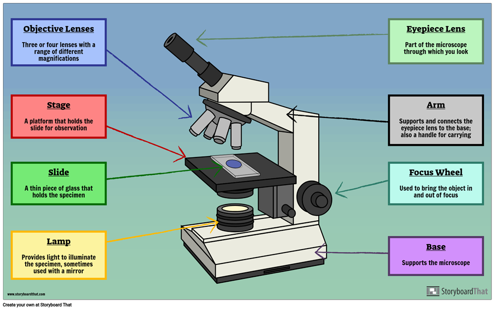
Parts of a microscope with functions and labeled diagram Microscope Definition Microscopes are instruments that are used in science laboratories to visualize very minute objects such as cells, and microorganisms, giving a contrasting image that is magnified. Microscopes are made up of lenses for magnification, each with its own magnification powers. Simple Microscope - Parts, Functions, Diagram and Labelling Parts of the optical parts are as follows: Mirror - A simple microscope has a plano-convex mirror and its primary function is to focus the surrounding light on the object being examined. Lens - The biconvex lens is placed above the stage and its function is to magnify the size of the object being examined.
Parts of Stereo Microscope (Dissecting microscope) - labeled diagram ... Labeled part diagram of a stereo microscope Major structural parts of a stereo microscope Optical components of a stereo microscope - definition and function Eyepieces Eyepiece tube Diopter adjustment ring Interpupillary Adjustment Objective Lenses Barlow lens Adjustment Knobs Light sources Stage plate Stage chips

Microscope diagram with labels and definitions
puzzles.mit.edu › 2016 › puzzleThe World's Longest Diagramless - MIT The World's Longest Diagramless Everything's bigger in Texas. In this diagramless crossword, Acrosses and downs have been merged into a single combined clue list in order of appearance. › 44090147 › CambridgeCambridge International AS and A Level Biology Coursebook ... Enter the email address you signed up with and we'll email you a reset link. Compound Microscope- Definition, Labeled Diagram, Principle, Parts, Uses The term "compound" in compound microscopes refers to the microscope having more than one lens. Devised with a system of combination of lenses, a compound microscope consists of two optical parts, namely the objective lens and the ocular lens. Working Principle of the Compound Microscope
Microscope diagram with labels and definitions. Compound Microscope Parts, Functions, and Labeled Diagram Compound Microscope Parts, Functions, and Labeled Diagram Parts of a Compound Microscope Each part of the compound microscope serves its own unique function, with each being important to the function of the scope as a whole. charlotte-engmann.de › magnification-quizEmail this Story to a Friend - charlotte-engmann.de 1 day ago · The microscope has been used in science to understand elements, diseases, and cells. Have a go at our magnification quiz! Look at the close-up . That depends partly on Mother Nature. Test Your Loupes & Magnification Knowledge. Sharing the microscope Often in labs you have to share the microscope with up to 3 other people. Microscope- Definition, Parts, Functions, Types, Diagram, Uses A microscope is an optical instrument having one or more lenses system which is used to get a clear magnified image of minute objects or structures that can't be viewed by the naked eyes. Derived from Greek words "mikrós " meaning "small" and "skópéō" meaning "look at " . They are devices used to observe the detailed structure of small objects. › articles › s41596/021/00556-8CODEX multiplexed tissue imaging with DNA-conjugated ... Jul 02, 2021 · A newly adapted version of CODEX uses an automated microfluidics system and conventional fluorescent microscope to iteratively hybridize, image and strip fluorescently labeled DNA probes that are ...
Metalloid - Wikipedia A metalloid is a type of chemical element which has a preponderance of properties in between, or that are a mixture of, those of metals and nonmetals.There is no standard definition of a metalloid and no complete agreement on which elements are metalloids. Despite the lack of specificity, the term remains in use in the literature of chemistry.. The six commonly recognised … Simple Microscope - Diagram (Parts labelled), Principle, Formula and Uses A simple microscope consists of Optical parts Mechanical parts Labeled Diagram of simple microscope parts Optical parts The optical parts of a simple microscope include Lens Mirror Eyepiece Lens A simple microscope uses biconvex lens to magnify the image of a specimen under focus. University of South Carolina on Instagram: “Do you know a future ... Oct 13, 2020 · 2,458 Likes, 119 Comments - University of South Carolina (@uofsc) on Instagram: “Do you know a future Gamecock thinking about #GoingGarnet? 🎉 ••• … Microscope Labeling Practice Diagram | Quizlet Start studying Microscope Labeling Practice. Learn vocabulary, terms, and more with flashcards, games, and other study tools.
Label Microscope Diagram - EnchantedLearning.com inclination joint - an adjustable joint that lets the arm tilt at various angles. low-power objective - a small lens with low magnifying power. mirror (or light source) - this directs light upwards onto the slide. revolving nosepiece - the rotating device that holds the objectives (lenses). stage - the platform on which a slide is placed. PDF Parts of a Microscope Printables - Homeschool Creations Label the parts of the microscope. You can use the word bank below to fill in the blanks or cut and paste the words at the bottom. Microscope Created by Jolanthe @ HomeschoolCreations.net. Parts of a eyepiece arm stageclips nosepiece focusing knobs illuminator stage objective lenses Laboratory procedures for diagnosis of anthrax, and isolation and ... 1. Anthrax and the microbiology laboratory; operational safety. With some country-to-country variation in safety level definitions and requirements, recommendations for the manipulation of the causative agent of anthrax, Bacillus anthracis, generally are that BSL (biosafety level) 2 practices, containment equipment and facilities are appropriate for diagnostic tests, but BSL3 … A Study of the Microscope and its Functions With a Labeled Diagram To better understand the structure and function of a microscope, we need to take a look at the labeled microscope diagrams of the compound and electron microscope. These diagrams clearly explain the functioning of the microscopes along with their respective parts. Man's curiosity has led to great inventions. The microscope is one of them.
Minds Eye: August 2015
Microscope labeled diagram - SlideShare Microscope labeled diagram 1. The Microscope Image courtesy of: Microscopehelp.com Basic rules to using the microscope 1. You should always carry a microscope with two hands, one on the arm and the other under the base. 2. You should always start on the lowest power objective lens and should always leave the microscope on the low power lens ...

Microscope Unlabelled Diagram - Micropedia
Microscope Parts, Function, & Labeled Diagram - slidingmotion Microscope parts labeled diagram gives us all the information about its parts and their position in the microscope. Microscope Parts Labeled Diagram The principle of the Microscope gives you an exact reason to use it. It works on the 3 principles. Magnification Resolving Power Numerical Aperture. Parts of Microscope Head Base Arm Eyepiece Lens
8.1 & 8.2 Math and Science: Onion Skin Lab
openstax.org › books › physics16.3 Lenses - Physics | OpenStax The ray diagram in Figure 16.33 shows image formation by the cornea and lens of the eye. The rays bend according to the refractive indices provided in Table 16.4 . The cornea provides about two-thirds of the magnification of the eye because the speed of light changes considerably while traveling from air into the cornea.

The Microscope: Create a Labelled Diagram | Teaching Resources
byjus.com There are also microscope types that find application in metallurgy and studying three-dimensional samples. In this article, there are 5 such microscope types that are discussed along with their diagram, working principle and applications. These five types of microscopes are: Simple microscope. Compound microscope.

Honors Biology Midterm Flashcards | Easy Notecards
Microscope Parts and Functions With Labeled Diagram and Functions How ... First, the purpose of a microscope is to magnify a small object or to magnify the fine details of a larger object in order to examine minute specimens that cannot be seen by the naked eye. Here are the important compound microscope parts... Eyepiece: The lens the viewer looks through to see the specimen.

The Animal Cell; What is the composition and functionality? by Ben Lieberman | Animal cell ...
(PDF) University Physics Volume1-OP - Academia.edu Enter the email address you signed up with and we'll email you a reset link.
Labelled Microscope with Functions Storyboard Szerint oliversmith
The World's Longest Diagramless - MIT The World's Longest Diagramless Everything's bigger in Texas. In this diagramless crossword, Acrosses and downs have been merged into a single combined clue list in order of appearance.
Plant Cell Anatomy - EnchantedLearning.com
Microscope: Types of Microscope, Parts, Uses, Diagram - Embibe Microscope: Definition, Parts, Parameters, Types, & Uses A microscope is an instrument that produces enlarged images of small objects. It provides the observer an exceedingly close view of minute structures at a scale convenient for examination and analysis. The microscope magnifies microscopic objects that are not visible to the naked eyes.

PZ C: compound microscope
Label the microscope — Science Learning Hub In this interactive, you can label the different parts of a microscope. Use this with the Microscope parts activity to help students identify and label the main parts of a microscope and then describe their functions. Drag and drop the text labels onto the microscope diagram.

All Saints Online: Diagram for Labelling: Microscope
Microscope, Microscope Parts, Labeled Diagram, and Functions Microscope, Microscope Parts, Labeled Diagram, and Functions What is Microscope? A microscope is a laboratory instrument used to examine objects that are too small to be seen by the naked eye. It is derived from Ancient Greek words and composed of mikrós, "small" and skopeîn,"to look" or "see".

PPT - Plant Cell Journal - Elodea PowerPoint Presentation - ID:1159196
Labeling the Parts of the Microscope | Microscope World Resources Labeling the Parts of the Microscope This activity has been designed for use in homes and schools. Each microscope layout (both blank and the version with answers) are available as PDF downloads. You can view a more in-depth review of each part of the microscope here. Download the Label the Parts of the Microscope PDF printable version here.

SEM and TEM Images - Plant Cells vs. Animal Cells
› books › NBK310485Laboratory procedures for diagnosis of anthrax, and isolation ... 1. Anthrax and the microbiology laboratory; operational safety. With some country-to-country variation in safety level definitions and requirements, recommendations for the manipulation of the causative agent of anthrax, Bacillus anthracis, generally are that BSL (biosafety level) 2 practices, containment equipment and facilities are appropriate for diagnostic tests, but BSL3 standards should ...

29 best Anatomy and Physiology images on Pinterest | Physiology, Anatomy and Anatomy reference
aoac-india.org › e-learningE-Learning – AOAC India Bright Field Microscope, Dark Field Microscope, Phase Contrast Microscope, Fluorescence Microscope, Confocal microscopy, Scanning and Transmission Electron Microscope and applications: 17th June 2019, 2019 11.30-12.30 pm: Watch Video: 50 : Nuclear magnetic resonance (NMR) – Part 1 DR. CHANDRASHEKHAR MR. RAGHAV MAVINKURVE, BRUKER
14 Best Images of Labeled Plant Cell Parts Worksheet - Prokaryotic Cell Coloring Page, Label ...
Inorganic Chemistry 4th edition, Catherine Housecroft Inorganic Chemistry 4th edition, Catherine Housecroft
Post a Comment for "38 microscope diagram with labels and definitions"